File:Figure 1. Fetal Lip and Primary Palate Three dimensional versus Two dimensional US.gif
Figure_1._Fetal_Lip_and_Primary_Palate_Three_dimensional_versus_Two_dimensional_US.gif (368 × 440 pixels, file size: 129 KB, MIME type: image/gif)
Figure 1.
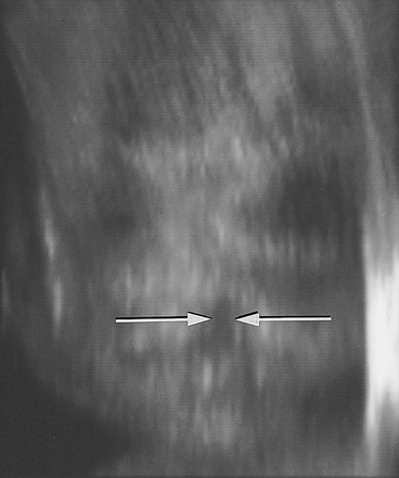
Three-dimensional rendered US image viewed frontally shows a facial cleft (arrows) in a fetus at 22 weeks gestational age. After viewing the rotating 3D image, the family elected to continue the pregnancy.[1]
Dear Maqdad Alsaif: The Radiological Society of North America (RSNA®) is pleased to grant you permission to reproduce the following figures in web format for use in your educational websites http://embryology.med.unsw.edu.au/embryology/index.php?title=Main_Page and http://embryology.med.unsw.edu.au/embryology/index.php?title=2011_Group_Project_11, provided you give full credit to the authors of the original publication.
Figures 1, 2, 3
Johnson D D, Pretorius D H, Budorick N E, et al. Fetal lip and primary palate: three-dimensional versus two-dimensional US. Radiology 2000;217:236-239.
Reference
<pubmed>11012450</pubmed>| [1]
- Note - This image was originally uploaded as part of a student project and may contain inaccuracies in either description or acknowledgements. Students have been advised in writing concerning the reuse of content and may accidentally have misunderstood the original terms of use. If image reuse on this non-commercial educational site infringes your existing copyright, please contact the site editor for immediate removal.
Cite this page: Hill, M.A. (2024, April 18) Embryology Figure 1. Fetal Lip and Primary Palate Three dimensional versus Two dimensional US.gif. Retrieved from https://embryology.med.unsw.edu.au/embryology/index.php/File:Figure_1._Fetal_Lip_and_Primary_Palate_Three_dimensional_versus_Two_dimensional_US.gif
- © Dr Mark Hill 2024, UNSW Embryology ISBN: 978 0 7334 2609 4 - UNSW CRICOS Provider Code No. 00098G
Assessment
+ US image of cleft is relevant to project. - what does "Figures 1, 2, 3" mean in the legend? - File name is not appropriate. It is not "Figure 1." of the project and should not have that included in the file name and "US" acronym should not be used. - This is in fact a 2 dimensional ultrasound image.
Overall ultrasound image is very relevant to the group project, this particular one is not the clearest and you have not attempted to explain the image correctly.
- ↑ <pubmed>11012450</pubmed>
File history
Click on a date/time to view the file as it appeared at that time.
| Date/Time | Thumbnail | Dimensions | User | Comment | |
|---|---|---|---|---|---|
| current | 21:40, 8 October 2011 |  | 368 × 440 (129 KB) | Z3284061 (talk | contribs) | Figure 1. Three-dimensional rendered US image viewed frontally shows a facial cleft (arrows) in a fetus at 22 weeks gestational age. After viewing the rotating 3D image, the family elected to continue the pregnancy.<ref name="PMID11012450"><pubmed>110124 |
| 20:17, 8 October 2011 |  | 368 × 440 (129 KB) | Z3284061 (talk | contribs) | Figure 1. Three-dimensional rendered US image viewed frontally shows a facial cleft (arrows) in a fetus at 22 weeks gestational age. After viewing the rotating 3D image, the family elected to continue the pregnancy.<ref name="PMID11012450"><pubmed>110124 |
You cannot overwrite this file.
File usage
The following 3 pages use this file:
