File:Fetal thymus.jpg
Fetal_thymus.jpg (450 × 600 pixels, file size: 122 KB, MIME type: image/jpeg)
Fetal Thymus
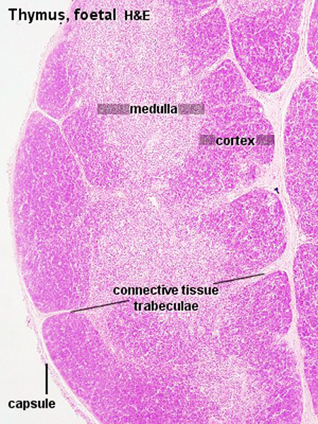
Thymus lymphoid tissue, overview 20th week of gestation.
- thymus is enclosed by a thin connective tissue capsule
- numerous septa extend into the thymus subdividing the two lobes
- two lobes divided into into numerous lobules (about 0.5 -2 mm in diameter).
- Blood vessels enter and leave the thymus via the connective tissue septa.
- Each lobulus is divided into
- Fetal Thymus Cortex - a darker peripheral zone.
- Fetal Thymus Medulla - a lighter central zone.
- Medullary tissue is continuous from lobule to lobule throughout each lobe.
See Unlabeled overview and also compare this structure with the Adult Thymus.
- Thymus Histology: Fetal Thymus overview | Fetal Thymus Medulla | Fetal Thymus Cortex | Adult Thymus | unlabeled fetal overview | unlabeled fetal medulla |unlabeled fetal thymic corpuscle |unlabeled fetal cortex | unlabeled adult overview | Category:Thymus | Immune System Development
Links: Histology | Histology Stains | Blue Histology images copyright Lutz Slomianka 1998-2009. The literary and artistic works on the original Blue Histology website may be reproduced, adapted, published and distributed for non-commercial purposes. See also the page Histology Stains.
Cite this page: Hill, M.A. (2024, April 19) Embryology Fetal thymus.jpg. Retrieved from https://embryology.med.unsw.edu.au/embryology/index.php/File:Fetal_thymus.jpg
- © Dr Mark Hill 2024, UNSW Embryology ISBN: 978 0 7334 2609 4 - UNSW CRICOS Provider Code No. 00098G
File history
Click on a date/time to view the file as it appeared at that time.
| Date/Time | Thumbnail | Dimensions | User | Comment | |
|---|---|---|---|---|---|
| current | 18:40, 25 February 2012 | | 450 × 600 (122 KB) | Z8600021 (talk | contribs) | increase image size |
| 11:01, 24 December 2010 | 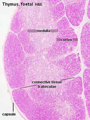 | 300 × 400 (28 KB) | S8600021 (talk | contribs) | Tyf04he.jpg |
You cannot overwrite this file.
File usage
The following 5 pages use this file: