File:Female- OHVIRA syndrome 01.jpg
Original file (340 × 1,000 pixels, file size: 76 KB, MIME type: image/jpeg)
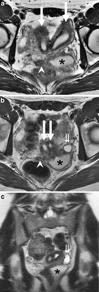
OHVIRA syndrome
Magnetic resonance image showing uterine didelphys, obstructed hemivagina, and ectopic ureter on MR imaging in a 17-year-old girl.
a Axial T2-W image demonstrates two widely separate uterine horns (large arrows), an obstructed left hemivagina distended with fluid (asterisk), and a nondilated right hemivagina (arrowhead).
b Axial T2-weighted image demonstrates two cervices (large arrows), an obstructed left hemivagina distended with fluid (asterisk), a nondilated right hemivagina (arrowhead), and a dilated left ureter (small arrows). On T1-W images, the fluid in the obstructed left hemivagina and the dilated left ureter was hypointense (not shown).
c Coronal T2-W image demonstrates the dilated left ureter (small arrows) inserting ectopically into the obstructed left hemivagina (asterisk). There is absence of visible left renal tissue (not shown)
Original file name: Fig. 1 http://www.ncbi.nlm.nih.gov/pmc/articles/PMC2817805/figure/Fig1/
Reference
<pubmed>19924410</pubmed>| PMC2817805
Pediatr Radiol. 2010 March; 40(3): 358–360.
Published online 2009 November 19. doi: 10.1007/s00247-009-1454-8.
Copyright © The Author(s) 2009
File history
Click on a date/time to view the file as it appeared at that time.
| Date/Time | Thumbnail | Dimensions | User | Comment | |
|---|---|---|---|---|---|
| current | 07:56, 30 October 2010 | 340 × 1,000 (76 KB) | S8600021 (talk | contribs) | ==OHVIRA syndrome== Uterine didelphys, obstructed hemivagina, and ectopic ureter on MR imaging in a 17-year-old girl. a Axial T2-W image demonstrates two widely separate uterine horns (large arrows), an obstructed left hemivagina distended with fluid (a |
You cannot overwrite this file.
File usage
The following page uses this file: