File:Establishment of HD hybrid cell line.jpg
Establishment_of_HD_hybrid_cell_line.jpg (600 × 600 pixels, file size: 93 KB, MIME type: image/jpeg)
Establishment of HD hybrid cell line
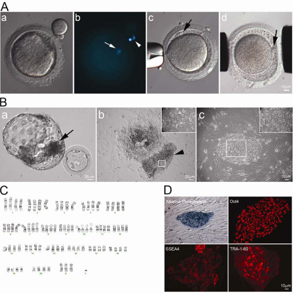
(A) First polar body of mature rhesus macaque oocyte was removed by gentle squeezing through a slit of zona pellucida (A-a). Staining of 1st polar body DNA (arrowhead) and oocyte DNA (arrow) (A-b). HD monkey skin cell was placed under the zona pellucida (black arrow) (A-c). Reconstructed oocyte with HD monkey skin cell (A-d; yellow arrow) was placed between two electrodes for electrofusion (A-d).
(B) Day 12 hatching blastocyst derived from HD monkey hybrid embryo (B-a; arrow indicated ICM). HD monkey hybrid blastocyst outgrowth at six days after attached onto feeder cells (B-b). High magnification of selected region (inset) of the ICM outgrowth (arrowhead). HD monkey hybrid cell line (TrES1) at passage 10 (B-c).
(C) G-banding analysis of TrES1. Cytogenetic analysis of TrES1 demonstrated tetraploid chromosome (84; XXXY).
(D) Expression of ES-cell specific markers: Alkaline phosphatase, Oct4, SSEA4 and TRA-1-60.
Reference
http://www.ncbi.nlm.nih.gov/pmc/articles/PMC2833146/?tool=pmcentrez
Copyright
This is an Open Access article distributed under the terms of the Creative Commons Attribution License (http://creativecommons.org/licenses/by/2.0), which permits unrestricted use, distribution, and reproduction in any medium, provided the original work is properly cited.
File history
Click on a date/time to view the file as it appeared at that time.
| Date/Time | Thumbnail | Dimensions | User | Comment | |
|---|---|---|---|---|---|
| current | 21:09, 17 August 2011 | | 600 × 600 (93 KB) | Z3290270 (talk | contribs) | (A) First polar body of mature rhesus macaque oocyte was removed by gentle squeezing through a slit of zona pellucida (A-a). Staining of 1st polar body DNA (arrowhead) and oocyte DNA (arrow) (A-b). HD monkey skin cell was placed under the zona pellucida ( |
You cannot overwrite this file.
File usage
The following 2 pages use this file: