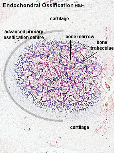
File:Endochondral ossification 1.jpg
From Embryology
No higher resolution available.
Endochondral_ossification_1.jpg (400 × 534 pixels, file size: 91 KB, MIME type: image/jpeg)
Endochondral ossification
- Bone Histology: Cartilage Histology | Histology Stains | Histology | cartilage | bone | bone timeline
- Trabecular bone trabecular | lamellar | trabecular - overview HE | trabecular - low HE | trabecular - med HE
- Endochondral ossification primary ossification | endochondral ossification
- Intramembranous ossification intramembranous - VG low | intramembranous - VG high | intramembranous - HE low | intramembranous - HE high
Links: Histology | Histology Stains | Blue Histology images copyright Lutz Slomianka 1998-2009. The literary and artistic works on the original Blue Histology website may be reproduced, adapted, published and distributed for non-commercial purposes. See also the page Histology Stains.
Cite this page: Hill, M.A. (2024, April 18) Embryology Endochondral ossification 1.jpg. Retrieved from https://embryology.med.unsw.edu.au/embryology/index.php/File:Endochondral_ossification_1.jpg
- © Dr Mark Hill 2024, UNSW Embryology ISBN: 978 0 7334 2609 4 - UNSW CRICOS Provider Code No. 00098G
Original file name: enos02he.jpg
File history
Click on a date/time to view the file as it appeared at that time.
| Date/Time | Thumbnail | Dimensions | User | Comment | |
|---|---|---|---|---|---|
| current | 17:15, 18 February 2013 | | 400 × 534 (91 KB) | Z8600021 (talk | contribs) |
You cannot overwrite this file.
File usage
The following page uses this file: