File:Embryo renal venous cartoon.jpg
From Embryology
No higher resolution available.
Embryo_renal_venous_cartoon.jpg (600 × 600 pixels, file size: 68 KB, MIME type: image/jpeg)
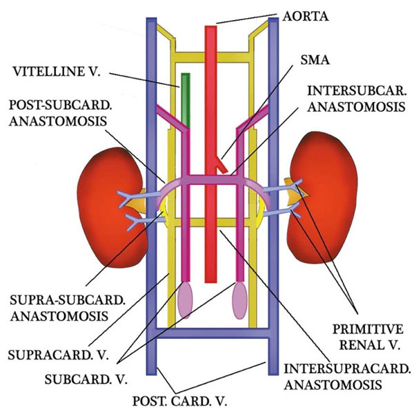
Embryo Renal Venous Cartoon
- Three pairs of veins appear in succession with regression of some portions and persistence of others.
- posterior cardinal → subcardinal → supracardinal veins
- Renal collar is formed by intersupracardinal anastomosis dorsally, intersubcardinal anastomosis ventrally and supra-subcardinal anastomosis laterally.
- Primitive dorsal and ventral renal veins drain into supra-subcardinal anastomoses.
- Both dorsal renal veins usually regress.
Reference
<pubmed>20461189</pubmed>| PMC2864862 | Korean J Radiol
This is an Open Access article distributed under the terms of the Creative Commons Attribution Non-Commercial License (http://creativecommons.org/licenses/by-nc/3.0) which permits unrestricted non-commercial use, distribution, and reproduction in any medium, provided the original work is properly cited.
File history
Click on a date/time to view the file as it appeared at that time.
| Date/Time | Thumbnail | Dimensions | User | Comment | |
|---|---|---|---|---|---|
| current | 13:14, 3 September 2011 | | 600 × 600 (68 KB) | S8600021 (talk | contribs) | ==Embryo Renal Venous Cartoon== * Three pairs of veins (posterior cardinal → subcardinal → supracardinal veins) appear in succession with regression of some portions and persistence of others. * Renal collar is formed by intersupracardinal anastomos |
You cannot overwrite this file.
File usage
The following 4 pages use this file: