File:Embryo-membranes stage 11.jpg
From Embryology
No higher resolution available.
Embryo-membranes_stage_11.jpg (600 × 568 pixels, file size: 61 KB, MIME type: image/jpeg)
Early Placentation (Carnegie stage 11)
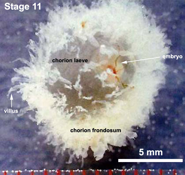
Embryo with intact placental membranes Carnegie stage 11 in Week 4.
Note this view is from the chorion laeve side with the chorion frondosum on the lower side. The thinness of the chorionic membrane allows the embryo to be seen inside the chorionic sac. Compare this week 4 with the a similar lateral view in week 7.
Description
- Embryo - Week 4, 23 - 26 days, 2.5 - 4.5 mm, Somite Number 13 - 20 (Scale 1 mm)
- The image shows the intact gestational sac (10 mm diameter) with chorionic villi (white finger-like extensions) covering the chorionic membrane surface.
- The chorion surface is now divided into 2 halves:
- chorion laeve - (laeve = smooth) The smooth chorion found on conceptus away from maternal blood supply (towards uterine epithelium and cavity) with very few villi present.
- chorion frondosum - (frondosum = leafy) the chorion found on conceptus oriented towards maternal blood supply where the majority of villi form and proliferate, will contribute the fetal component of the future placenta.
Cite this page: Hill, M.A. (2024, April 20) Embryology Embryo-membranes stage 11.jpg. Retrieved from https://embryology.med.unsw.edu.au/embryology/index.php/File:Embryo-membranes_stage_11.jpg
- © Dr Mark Hill 2024, UNSW Embryology ISBN: 978 0 7334 2609 4 - UNSW CRICOS Provider Code No. 00098G
File history
Click on a date/time to view the file as it appeared at that time.
| Date/Time | Thumbnail | Dimensions | User | Comment | |
|---|---|---|---|---|---|
| current | 11:17, 6 October 2010 | | 600 × 568 (61 KB) | S8600021 (talk | contribs) | ==Embryo with intact placental membranes (Carnegie stage 11)== '''Links:''' Placenta Development {{Template:UNSWimages}} Category:Placenta Category:Human Embryo Category:Carnegie Stage 11 |
You cannot overwrite this file.