File:Early human telomeres.jpg
Original file (1,280 × 1,006 pixels, file size: 194 KB, MIME type: image/jpeg)
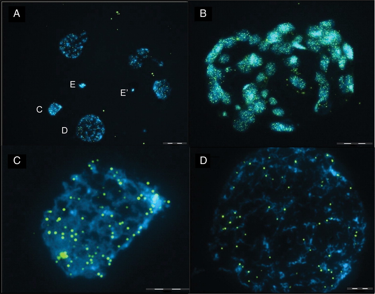
Early Human Telomeres
Telomeres in human embryos. Telomeres were stained using a FITC-labelled pan-telomeric probe (green). All nuclei were counterstained with DAPI (blue).
(A) 6-cell embryo resulting in five nuclei with two polar bodies, E and E′ (scale bar = 44 µm).
Two of the nuclei (C and D, scale bar = 8.5 µm) are also shown under higher magnification (scale bar = 4 µm).
(B) The telomeres in a fixed blastocyst (scale bar = 55 µm).
- "Telomeres are repeated sequences that protect the ends of chromosomes and harbour DNA repair proteins. Telomeres shorten during each cell division in the absence of telomerase. When telomere length becomes critically short, cell senescence occurs. Telomere length therefore reflects both cellular ageing and capacity for division. We have measured telomere length in human germinal vesicle (GV) oocytes and preimplantation embryos, by quantitative fluorescence in situ hybridization (Q-FISH), providing baseline data towards our hypothesis that telomere length is a marker of embryo quality. The numbers of fluorescent foci suggest that extensive clustering of telomeres occurs in mature GV stage oocytes, and in preimplantation embryos. When calculating average telomere length by assuming that each signal presents one telomere, the calculated telomere length decreased from the oocyte to the cleavage stages, and increased between the cleavage stages and the blastocyst (11.12 versus 8.43 versus 12.22 kb, respectively, P < 0.001). Other methods of calculation, based upon expected maximum and minimum numbers of telomeres, confirm that telomere length in blastocysts is significantly longer than cleavage stages. Individual blastomeres within an embryo showed substantial variation in calculated average telomere length. This study implies that telomere length changes according to the stage of preimplantation embryo development."
Original File Name: Figure 1 F1.large.jpg http://molehr.oxfordjournals.org/content/16/9/685/F1.expansion.html
Reference
<pubmed>20573647</pubmed>| PMC2930518 | Mol Hum Reprod.
© The Author 2010. Published by Oxford University Press on behalf of the European Society of Human Reproduction and Embryology. This is an Open Access article distributed under the terms of the Creative Commons Attribution Non-Commercial License (http://creativecommons.org/licenses/by-nc/2.5), which permits unrestricted non-commercial use, distribution, and reproduction in any medium, provided the original work is properly cited.
File history
Click on a date/time to view the file as it appeared at that time.
| Date/Time | Thumbnail | Dimensions | User | Comment | |
|---|---|---|---|---|---|
| current | 11:05, 25 October 2010 | | 1,280 × 1,006 (194 KB) | S8600021 (talk | contribs) | ==Early Human Telomeres== Telomeres in human embryos. Telomeres were stained using a FITC-labelled pan-telomeric probe (green). All nuclei were counterstained with DAPI (blue). (A) 6-cell embryo resulting in five nuclei with two polar bodies, E and E′ |
You cannot overwrite this file.
File usage
The following 3 pages use this file: