File:DeBruin1910 fig10.jpg

Original file (1,422 × 1,100 pixels, file size: 208 KB, MIME type: image/jpeg)
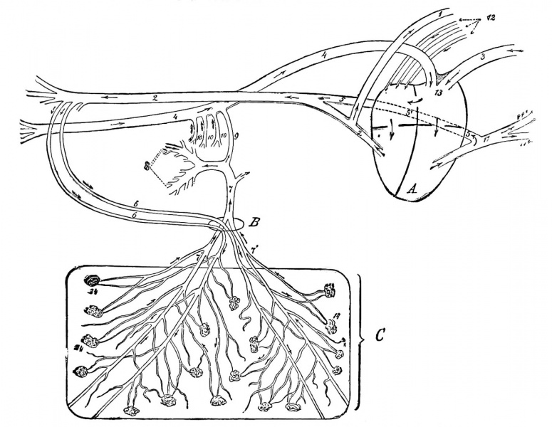
Fig. 10. Schematic Sketch of the Foetal Circulation of a Calf.
The arrows indicate the direction in which the blood flows.
A, Heart; B, umbilical opening; C, portion of the chorion. 1, Anterior aorta; 2, posterior aorta; 3 anterior vena cava; 4, posterior vena cava; 5, duct of Botalli; part of Botalli's duct posterior to the heart (sketched somewhat too long, but was necessary in order to demonstrate it) ; 6, umbilical arteries; 7, umbilical vein; 7', some of its branches; 8, portal vein; 9, ductus venosus; 10, portal veins: 11, pulmonary artery; 11', some of its branches; 12, pulmonary veins; 13, tuberculum Loweri; 14, chorion papillae.
- Links: Bovine Development | Placenta Development
| Historic Disclaimer - information about historic embryology pages |
|---|
 |
Fetal Circulation
This early circulation, which might be called the omphalomesenteric circulation {circulatio omphalo mesenterica), exists, same as the umbilical vesicle, only for a time. When the latter disappears, and the allantois forms, it is replaced by the placental circulation, representing the foetal circulation (Fig. 10). By means of this circulation, the blood which circulated in the foetus is taken to the foetal placenta and reaches the smaller vessel of the chorion. Here its composition experiences chauges and then returns to the foetus. In the placenta an exchange of oxygen for carbon dioxide occurs, the circulation at this part fulfilling the same purpose as the pulmonary circulation does in the lungs. The venous blood in the placenta gives off carbon and takes up oxygen. This process is the so-called placental respiration. The blood oxygenated in the placenta reaches the foetus at the umbilical ring by the two umbilical veins. Within the umbilical ringthe two veins coalesce to form one vessel, which leads to the liver {porta Jiepatica), first giving off a branch {ductus^ Arantii) emptying into the posterior vena cava. The remaining trunk forms one vessel with the portal vein. At this spot, then, the first mixture of the arterial blood of the umbilical veins with the hepatic venous blood takes place. The trunk of the umbilical veins divides in the liver into numerous capillaries, from -which finally the hepatic veins arise aiid empty into the posterior vena cava. The arterial blood of the umbilical veins, which, on account of its mixing with the vena portse, has been modified in its composition, now undergoes a second mixing, namely, with the blood of the posterior vena cava.
Only a part of the blood of the umbilical veins circulates through the liver, while another part, as already related, empties into the posterior vena cava, via the ductus Arantii. Hepatic veins aud ductus Arantii finally carry the umbilical veins' blood into the posterior vena cava, and the latter takes it to the left auricle of the heart. When the blood arrives in the right auricle, it runs through the oval foramen (situated in the septum between right and left auricle) into the left auricle, the blood current being guided by an eminence, the iubercidum Loweri.
During Foetal life an oval orifice {foramen ovale) is found replaced in the adult by the fossa ovalis. It has a diameter of 1 cm., has an infundibuliform opening into the right auricle, and guides the blood carried by the posterior vena cava directly into the left auricle. On that side of the septum turned toward the left auricle lies the funnel-shaped valve of the oval foramen {valvula foraminis ovalis), formed by a fold of the endocardium. It is attached to the edge of the oval foramen, and its perforated infundibulum projects into the left auricle. This valve prevents a return flow of the blood from the left into the right auricle. In the left auricle the blood coming from the right auricle mixes with the venous blood of the pulmonary veins. this mixture is of little moment, as the amount of blood flowing through the Inngs during intrauterine life is a small quantity. From the left auricle the Foetal blood goes through the auriculo-ventricular opening into the left auricle and on through the aorta to the various portions of the body. Blood mixtures take place at the following places :
- At the portal fissure, a confluence of the vena portse and arterial blood of the umbilical veins.
- At the end of the ductus Arantii, into the posterior vena cava, and at the end of the hepatic veins, also into the last named.
- In the left auricle, a mixing of the pulmonary blood, carried by the posterior vena cava from the right to the left auricle. The blood of the anterior aorta supplying the anterior extremity experiences now no further change. After coui'sing through the capillaries into the veins, it returns through the anterior vena cava to the right auricle. From here it gains the right ventricle and pulmonary artery. But the greater part flows through Botalli's duct {ductus Botalli) into the posterior aorta.
The ductus Botalli is a short tube running obliquely from left to right and before to behind, and connects the pulmonary artery v^itli the posterior aorta. A very small amount of the blood reaches the lungs via the pulmonary artery (arteria pidinonaris), and from here, after circulating through tjje capillaries, arrives in the left auricle by the left auricle.
The placenta is the organ of respiration. What the lungs are to the breathing animal the placenta is to the foetus. In the placenta, more than a simple exchange of carbon for oxygen takes place. In all probability, nutritive elements pass the placental filter, although apparently during this process a modification in the composition occurs. Between the villi of the foetal and maternal placenta a thin layer of uterine milk is found. One is of the opinion that nutritive elements from the capillaries of the uterine mucosa carried into the epithelium of the maternal placenta are there changed into uterine milk. Bonnet proved that the fat of the uterine inilk is no product of degeneration, but the result of infiltration.
The uterine milk, or certain of its constituents, under. the influence of pressure, are supposed to enter the epithelia of the villi of the chorion, and reach here the blood current of the foetus. According to this hypothesis, uterine milk plays as important a role in the later foetal periods as at the time when the various organs first begin to develop.
Experiments show that under high pressure white blood, cells, pigment and bacilli pass the placental filter; under ordinary conditions, with moderate pressure, this does not occur readily. The baccilus of glanders, tuberculosis, pass through that filter, but the anthrax bacillus does not invariably.
That tubercule bacilli penetrate the placental foetus, the various cases of tuberculosis of the bovine foetus reported in our literature prove (Bang, Johne, Korevaar, Lungwitz). In the case reported by Korevaar, no tuberculosis of the uterus was present.
DeBruin Bovine Obstetrics (1910)
Cite this page: Hill, M.A. (2024, April 20) Embryology DeBruin1910 fig10.jpg. Retrieved from https://embryology.med.unsw.edu.au/embryology/index.php/File:DeBruin1910_fig10.jpg
- © Dr Mark Hill 2024, UNSW Embryology ISBN: 978 0 7334 2609 4 - UNSW CRICOS Provider Code No. 00098G
File history
Click on a date/time to view the file as it appeared at that time.
| Date/Time | Thumbnail | Dimensions | User | Comment | |
|---|---|---|---|---|---|
| current | 03:49, 4 November 2013 | | 1,422 × 1,100 (208 KB) | Z8600021 (talk | contribs) | ==Fig. 10== {{Historic Disclaimer}} |
You cannot overwrite this file.
File usage
The following page uses this file: