File:Complete hydatidiform mole 02.jpg
Complete_hydatidiform_mole_02.jpg (748 × 560 pixels, file size: 60 KB, MIME type: image/jpeg)
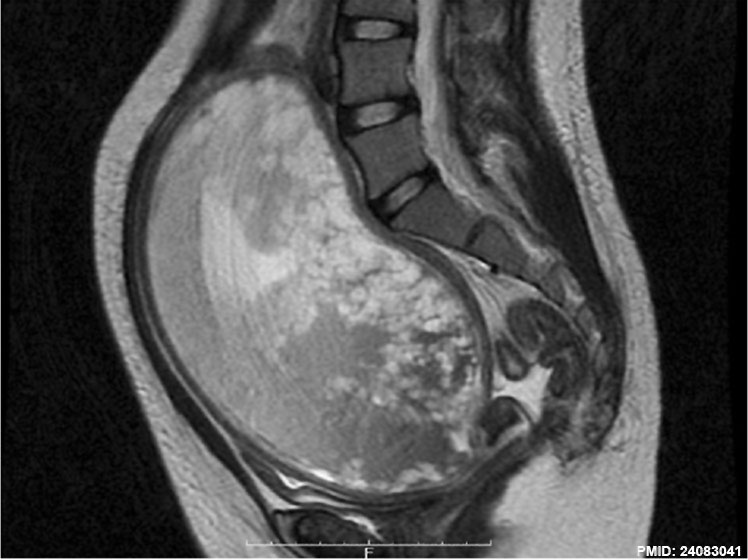
Complete Hydatidiform Mole
The magnetic resonance image (T2 weighted, sagittal) shows a massive intrauterine mass (19 × 15 × 10 cm) with many small vesicles, no normal gestational sac, and no fetus.
Complete Mole - Only paternal chromosomes, chromosomal genetic material from the oocyte (ovum or egg) is lost, by an unknown process.
- Hydatidiform Mole Links: Transvaginal ultrasound | Magnetic resonance image | Macroscopic image | Histology | Hydatidiform Mole
Reference
<pubmed>24083041</pubmed>| Case Rep Obstet Gynecol.
Copyright
© 2013 Naoki Matsumoto et al. This is an open access article distributed under the Creative Commons Attribution License, which permits unrestricted use, distribution, and reproduction in any medium, provided the original work is properly cited.
Figure 1 267268.fig.001a.jpg http://www.hindawi.com/journals/criog/2013/267268/fig1/ Image adjusted in size contrast and labelling.
Cite this page: Hill, M.A. (2024, April 25) Embryology Complete hydatidiform mole 02.jpg. Retrieved from https://embryology.med.unsw.edu.au/embryology/index.php/File:Complete_hydatidiform_mole_02.jpg
- © Dr Mark Hill 2024, UNSW Embryology ISBN: 978 0 7334 2609 4 - UNSW CRICOS Provider Code No. 00098G
File history
Click on a date/time to view the file as it appeared at that time.
| Date/Time | Thumbnail | Dimensions | User | Comment | |
|---|---|---|---|---|---|
| current | 14:52, 10 May 2014 | | 748 × 560 (60 KB) | Z8600021 (talk | contribs) | ==Complete Hydatidiform Mole== (a) Transvaginal ultrasonography (sagittal). (b) The magnetic resonance image (T2 weighted, sagittal) shows a massive intrauterine mass (19 × 15 × 10 cm) with many small vesicles, no normal gestational sac, a... |
You cannot overwrite this file.
File usage
The following 3 pages use this file: