File:Colon histology 008.jpg
From Embryology

Size of this preview: 799 × 600 pixels. Other resolution: 1,278 × 959 pixels.
Original file (1,278 × 959 pixels, file size: 237 KB, MIME type: image/jpeg)
Human Ano-Rectal Junction Histology
- alimentary canal, GIT, large intestine, rectum, mucosa, crypt of Lieberkühn, longitudinal section, AB/VG
- Stain: van Gieson
- Colon Histology Links: Ano-Rectal Junction Overview Labeled | Colon Wall Labeled | Colon Mucosa Labeled | Colon Overview | Ano-Rectal Junction Overview | Intestinal Gland - longitudinal van Gieson | Intestinal Gland - transverse van Gieson | Intestinal Gland - longitudinal H&E | Intestinal Gland - transverse H&E | GIT Histology | Gastrointestinal Tract Development
Original File Name: arec02vg.jpg
Links: Histology | Histology Stains | Blue Histology images copyright Lutz Slomianka 1998-2009. The literary and artistic works on the original Blue Histology website may be reproduced, adapted, published and distributed for non-commercial purposes. See also the page Histology Stains.
Cite this page: Hill, M.A. (2024, April 24) Embryology Colon histology 008.jpg. Retrieved from https://embryology.med.unsw.edu.au/embryology/index.php/File:Colon_histology_008.jpg
- © Dr Mark Hill 2024, UNSW Embryology ISBN: 978 0 7334 2609 4 - UNSW CRICOS Provider Code No. 00098G
File history
Click on a date/time to view the file as it appeared at that time.
| Date/Time | Thumbnail | Dimensions | User | Comment | |
|---|---|---|---|---|---|
| current | 11:24, 2 November 2009 | | 1,278 × 959 (237 KB) | S8600021 (talk | contribs) | |
| 11:23, 2 November 2009 | 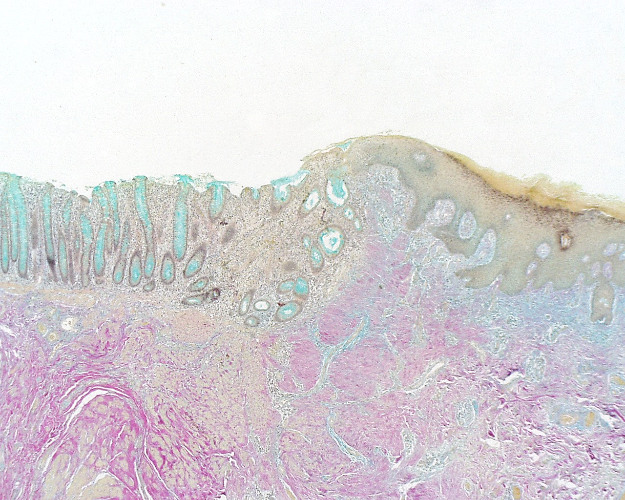 | 1,280 × 1,024 (461 KB) | S8600021 (talk | contribs) |
You cannot overwrite this file.
File usage
The following 3 pages use this file: