File:Cardiac muscle EM01.jpg

Original file (1,072 × 735 pixels, file size: 231 KB, MIME type: image/jpeg)
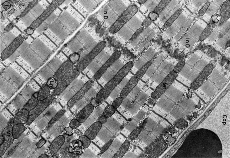
Cardiac Muscle Electron Micrograph
Electron micrograph of parts of three cat cardiac muscle fibers and an adjacent capillary in longitudinal section. This is a historic (1969) EM showing key features in cat cardiac ventricular muscle ultrastructure. Only the intercalated disc and some cross-striations can typically be seen in the light microscope histology slides.
Legend
- In D - Intercalated disc, the two lower cells are joined end to end by a typical steplike intercalated disc.
- Mt - Rows of mitochondria appear to divide the contractile substance into myofibril-like units but, unlike the true myofibrils of skeletal muscle, these branch and rejoin and are quite variable in width.
- Lp - Lipid droplets somewhat distorted in specimen preparation are found between the ends of the mitochondria.
- Cap - Capillary.
Original image X 15,000.
Reference
Fawcett DW & McNutt NS. (1969). The ultrastructure of the cat myocardium. I. Ventricular papillary muscle. J. Cell Biol. , 42, 1-45. PMID: 4891913
Copyright
Rockefeller University Press - Copyright Policy This article is distributed under the terms of an Attribution–Noncommercial–Share Alike–No Mirror Sites license for the first six months after the publication date (see http://www.jcb.org/misc/terms.shtml). After six months it is available under a Creative Commons License (Attribution–Noncommercial–Share Alike 4.0 Unported license, as described at https://creativecommons.org/licenses/by-nc-sa/4.0/ ). (More? Help:Copyright Tutorial)
Original article figure (FIG. 1) has been scaled and rotated.
Cite this page: Hill, M.A. (2024, April 25) Embryology Cardiac muscle EM01.jpg. Retrieved from https://embryology.med.unsw.edu.au/embryology/index.php/File:Cardiac_muscle_EM01.jpg
- © Dr Mark Hill 2024, UNSW Embryology ISBN: 978 0 7334 2609 4 - UNSW CRICOS Provider Code No. 00098G
File history
Click on a date/time to view the file as it appeared at that time.
| Date/Time | Thumbnail | Dimensions | User | Comment | |
|---|---|---|---|---|---|
| current | 11:52, 6 August 2012 | | 1,072 × 735 (231 KB) | Z8600021 (talk | contribs) | ==Cardiac Muscle EM== Electron micrograph of parts of three cat cardiac muscle fibers and an adjacent capillary (Cap) in longitudinal section. The two upper cells are joined end to end by a typical steplike intercalated disc (In D).Rows of mitochondria ( |
You cannot overwrite this file.
File usage
The following 5 pages use this file: