File:Cardiac Conduction System.jpg
From Embryology
Size of this preview: 800 × 574 pixels. Other resolution: 1,201 × 862 pixels.
Original file (1,201 × 862 pixels, file size: 81 KB, MIME type: image/jpeg)
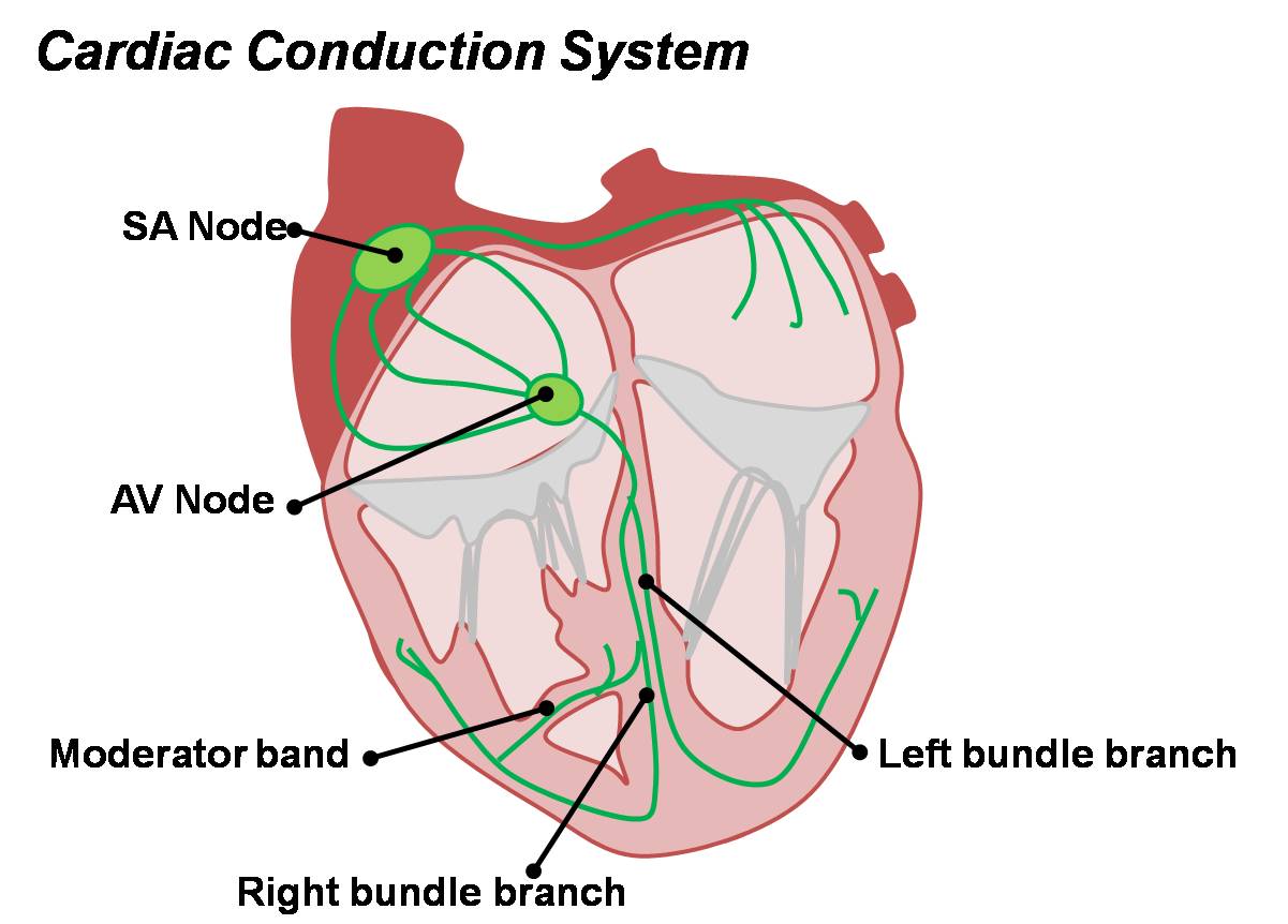
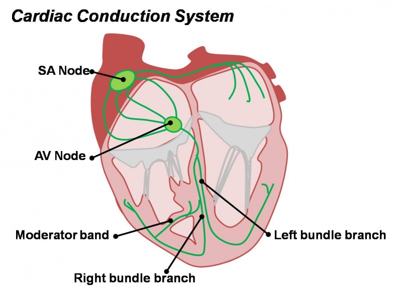
Cardiac Conduction System in the Adult Heart
Electrical conduction system of the heart occurs through specially modified cardiac muscle cells.
- Week 7 GA - SA node initially develops in the sinus venosus and then is incorporated into the RA.
- AV node arises slightly superior to the endocardial cushions.
- SA node - sinoatrial node located on the anterior border of the opening of the superior vena cava.
- AV node - atrioventricular node located near the orifice of the coronary sinus in the annular and septal fibres of the right atrium.
- bundle of His
- Left bundle branch
- Right bundle branch
- moderator band - primary conduction path in to the free wall originating from the right bundle branch.
| Begin Advanced | Heart Fields | Heart Tubes | Cardiac Looping | Cardiac Septation | Outflow Tract | Valve Development | Cardiac Conduction | Cardiac Abnormalities | Molecular Development |
| Cardiac Embryology | Begin Basic | Begin Intermediate | Begin Advanced |
References
<pubmed>19808465</pubmed> <pubmed>16148066</pubmed> <pubmed>20811536</pubmed> <pubmed>12382942</pubmed> <pubmed>20235167</pubmed>
Cite this page: Hill, M.A. (2024, April 19) Embryology Cardiac Conduction System.jpg. Retrieved from https://embryology.med.unsw.edu.au/embryology/index.php/File:Cardiac_Conduction_System.jpg
- © Dr Mark Hill 2024, UNSW Embryology ISBN: 978 0 7334 2609 4 - UNSW CRICOS Provider Code No. 00098G
File history
Click on a date/time to view the file as it appeared at that time.
| Date/Time | Thumbnail | Dimensions | User | Comment | |
|---|---|---|---|---|---|
| current | 12:03, 14 March 2010 | | 1,201 × 862 (81 KB) | Z3212774 (talk | contribs) | category:Heart ILP Cardiac conduction system in the adult heart. |
You cannot overwrite this file.