File:BrauneB1.jpg
Original file (1,200 × 485 pixels, file size: 139 KB, MIME type: image/jpeg)
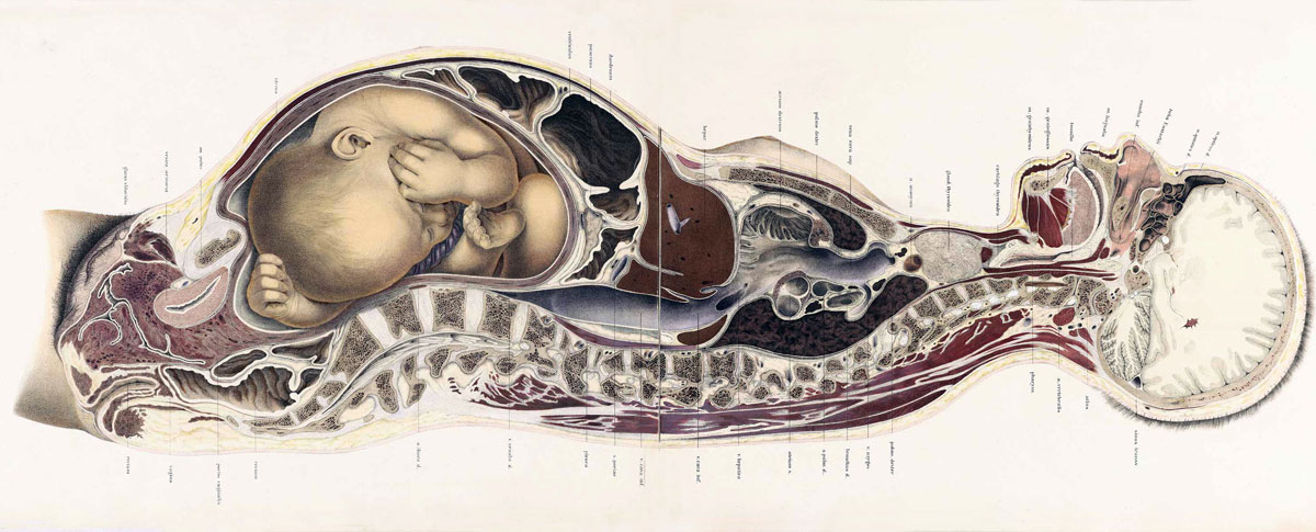
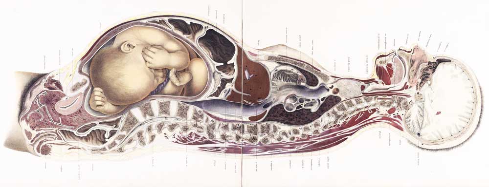
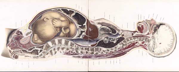
The Position of the Uterus and Fetus at Term (1872)
Braune, Wilhelm (1831-1892): Die Lage des Uterus und Foetus am Ende der Schwangerschaft. (Leipzig: Verlag von Viet & Comp., 1872).
This sagittal section through the maternal anatomy shows the relative size and position of the fetus during pregnancy.
Note also the altered maternal anatomical relationships during the pregnancy.
- Braune Image Links: Uterus and Fetus Position at Term | Section through Uterus and Fetus | Uterus without Fetus | Uterus and Fetus Position at Birth | Section through Uterus and Fetus at Birth | Uterus without Fetus at Birth | 17-18 C Anatomies
The body from which this preparation was made was quite recent, twenty-five years of age, in the last month of pregnancy, and received in the condition of rigor mortis. Beyond the constriction of the neck produced by the means of death (hanging) no abnormality existed. The condition of the genitals corresponded with an advanced stage of pregnancy, and were injected and succulent. The method of preparation was carried out in the usual manner.
| Preparation Method |
|---|
| The foetus, which was divided in the section of the body, was subsequently restored to its original condition, so as to afford a representation of its former position in the uterus. I chiselled out the foetus and the liquor amnii from the left side of the body, and moistened the surface of the section of the uterus, and then froze it on the right side. The portion now lying in the right half of the uterus remained then for the purposes of representation as an untouched foetus. The left half of the uterus and its appendages, after the removal of the rest of the liquor amnii, was represented as empty. The foetus, which was in the second position of the head, was a well-formed female. The vulva were closed and the nails well developed. Its entire length was about twenty-three inches, its weight without the cord about six pounds. The cord was divided, and passed to the placenta between the head and right arm, the placenta being placed downwards and on the right side of the uterus. The child, as the plate shows, lay mostly in the right half of the uterus. In the section more than the right half of the head which was sawn obliquely, was removed. Moreover, the left arm and a portion of the right shoulder were divided longitudinally, and the forearm being placed at right angles with it, transversely, as well as a portion of the right leg, which extended towards the left side. The left knee was moreover grazed by the saw. The back and belly lay in the right half of the uterus, and the greater portion of the liquor amnii remained in the left. As the relations of this oblique section of the foetus offer points of no peculiar interest, I have refrained from reproducing the corresponding plate of the large atlas in this small edition. |
The uterus is so folded over the symphysis that its anterior wall forms a kind of sac, indicating a condition of relaxation. The numerous large veins in its tissue are shown in the plate in the wall as simple strokes, their lumina becoming recognisable only when their walls were separated from each other; they appear patent, however, in the vaginal portion of the uterus and in the vagina itself. The vaginal portion of the uterus is proportionately deep, and for the most part lies in the left half of the body, the section having passed through its right half and opened merely the first portion of the cervix, as shown in Plate XXIX A. It was filled with viscid mucus and opened into the cavity of the uterus, about one fifth of an inch below the plane of section, so that its upper half could not be seen. The length of the vagina at this period of pregnancy makes it probable that the woman was not a primipara, notwithstanding that there were no cicatrices on the abdominal parietes, and the os internum was so narrow that only a very small sound could pass it. The number of veins met with in the right half of the vagina and their swollen condition is remarkable, and their lumina are peculiarly well seen in the left half of the preparation, Plate XXIX B. The falling in of the vaginal portion of the uterus is remarkable, considering the empty contracted condition of the bladder. The latter has slipped down bodily from the inner surface of the symphysis, and is so completely displaced that the course of the urethra has become bent at an angle. The external os lies in the hollow of the under border of the symphysis, although, according to Moreau, it corresponds at the end of pregnancy with the level of the upper border of the symphysis, and is still higher according to Schultze.
The level of the fundus corresponds nearly with the under border of the first lumbar vertebra; a more accurate definition cannot be given, as the highest point of the uterus was not included in the section, as it inclined more to the right side. This is almost the level given by Moreau, and according to the measurements of Schultze (' Wandtafeln,' taf. vi), it would appear to be the second lumbar vertebra. As the parts in the meanwhile began to thaw, a more accurate measurement in this particular could not be made.
The depth of the cavity of the uterus and its connections, and of the entire cavity of the abdomen, is less than is usually admitted. Notwithstanding the size of the foetus it is not improbable that the attitude of the body had some influence in this respect, and that lying horizontally on the back the uterus obtained a kind of fulcrum on the vertebral column, whilst in the upright position the yielding walls of the abdomen are pushed forwards. It is farther to be remembered that in dead bodies generally in consequence of the high position of the diaphragm, the depth of the cavity of the abdomen is less than during life.
In the present instance the distance of the lumbar vertebrae from the anterior wall of the abdomen was almost one third of the entire sagittal diameter of the body at its point of greatest distension ; whilst in the body which in Plate II is represented in the second month of pregnancy, the lumbar spine projects slightly beyond the middle of this diameter.
Finally, the vessels were in this case uninjected a circumstance which is to be taken into consideration in estimating the thickness of the walls of the uterus.
| Abdomen |
|---|
|
The cavity of the abdomen extended tolerably far up in comparison with its slight depth. The highest point of the diaphragm reached the level of the seventh dorsal vertebra, whilst in males, and unimpregnated females of middle age it would extend only as far as the ninth or tenth.
|
| Thorax |
|---|
|
The thoracic cavity appears shallow, in consequence of the high position of the diaphragm, but, on the other hand, very wide in the antero-posterior diameter, as may be seen by comparing this preparation with the section of the female subject in Plate II. But on the strength of this, an enlargement of the base of the thorax during pregnancy is not necessarily to be inferred, as measurements for comparison are wanting before and after it. Although it may appear plausible to explain the unvarying size of the spirometer during pregnancy, by the fact that the diminution of the thoracic space dependent on the rising of the diaphragm is compensated for by the traction of the abdominal muscles acting over the uterus like a pulley, the anatomical relations in this respect are not yet determined. Gerhard found, by measurements on living bodies, that in forty-two females in advanced pregnancy the diaphragm was in thirty-six cases in a normal position, in five it was deeper, and only in one higher. Dorn in his measurements by means of the cyrtometer on living females in advanced pregnancy and in lying-in women, found that in most cases the bases of the thorax had a greater breadth during pregnancy than after delivery, but, on the other hand, its depth was less from before backwards. When the uterus was empty this relation was reversed, the thorax collapsed on both sides, the transverse diameter decreased, and the vertical diameter increased (' Bericht iiber die Naturforchenversammlung zu Griessen,' 1865, p. 225).
|
The soft parts of the neck are considerably dislocated towards the left side, owing to the hypertrophied thyroid body. The trachea lies so far over to the left side that only a small portion of the thyroid cartilage is met with.
The brain was divided through its right half, the radiation of the fibres of the right corpus callosum being thus shown. Beneath it is the descending cornu of the right lateral ventricle with the pes hippocampi. Beneath the dura mater, in the right half of the preparation, a portion of the Gasserian ganglion and some fibres of the fifth nerve are seen.
| Skeleton |
|---|
The question arises whether, in a weak obliquely contracted pelvis, showing such a variation, child-birth be possible without surgical aid.
Detailed description of this Plate XXIX A, XXIX B, and XXX | Uterus and Fetus Position at Term - original version
Reference
Braune W. An atlas of topographical anatomy after plane sections of frozen bodies. (1877) Trans. by Edward Bellamy. Philadelphia: Lindsay and Blakiston.
http://www.nlm.nih.gov/exhibition/historicalanatomies/braune_home.html
- NLM Copyright Information: Government information at NLM Web sites is in the public domain. Public domain information may be freely distributed and copied, but it is requested that in any subsequent use the National Library of Medicine (NLM) be given appropriate acknowledgement. When using NLM Web sites, you may encounter documents, illustrations, photographs, or other information resources contributed or licensed by private individuals, companies, or organizations that may be protected by U.S. and foreign copyright laws. Transmission or reproduction of protected items beyond that allowed by fair use as defined in the copyright laws requires the written permission of the copyright owners. Specific NLM Web sites containing protected information provide additional notification of conditions associated with its use.
Cite this page: Hill, M.A. (2024, April 18) Embryology BrauneB1.jpg. Retrieved from https://embryology.med.unsw.edu.au/embryology/index.php/File:BrauneB1.jpg
- © Dr Mark Hill 2024, UNSW Embryology ISBN: 978 0 7334 2609 4 - UNSW CRICOS Provider Code No. 00098G
File history
Click on a date/time to view the file as it appeared at that time.
| Date/Time | Thumbnail | Dimensions | User | Comment | |
|---|---|---|---|---|---|
| current | 15:34, 30 October 2012 | 1,200 × 485 (139 KB) | Z8600021 (talk | contribs) | ||
| 18:31, 10 September 2009 | 1,000 × 384 (48 KB) | S8600021 (talk | contribs) | |||
| 15:09, 22 July 2009 | 600 × 241 (20 KB) | MarkHill (talk | contribs) | Braune, Wilhelm (1831-1892): Topographisch-anatomischer Atlas : nach Durchschnitten an gefrornen Cadavern, Leipzig: Verlag von Veit & Comp., 1867-1872. (Topographic-anatomical Atlas) http://www.nlm.nih.gov/exhibition/historicalanatomies/braune_home.html |
You cannot overwrite this file.
File usage
The following 21 pages use this file:
- 2009 Lecture 1
- 2010 Lecture 1
- ANZACA Meeting 2012 - Embryology
- BGDA Practical 12 - Birth
- BGDA Practical 12 - Third Trimester
- Book - An Atlas of Topographical Anatomy (1877)
- Book - An Atlas of Topographical Anatomy 30
- Embryology History
- Embryology History - 17th and 18th Century Anatomies
- Fetal Development
- Lecture - 2011 Course Introduction
- Lecture - 2014 Course Introduction
- Lecture - 2015 Course Introduction
- Lecture - 2016 Course Introduction
- Lecture - 2017 Course Introduction
- Maternal Development
- Museum of Natural History Berlin - 2013 Seminar
- Talk:Lecture - 2016 Course Introduction
- Template talk:Braune images
- Special:Badtitle/NS501:Embryology History
- History:Embryology History