File:Bone marrow histology 04.jpg
Bone_marrow_histology_04.jpg (480 × 600 pixels, file size: 61 KB, MIME type: image/jpeg)
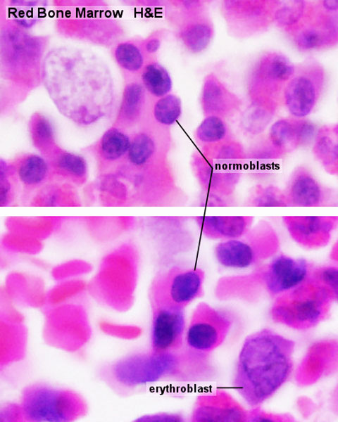
Bone Marrow Histology
Erythropoiesis is the proerythroblast - a large, slightly basophilic cell, which contains a large, lightly stained nulceus.
Proerythroblast
Proerythroblasts proliferate to generate a sequence of cells which show a gradual decrease in size and condensation of their chromatin. named after changes in the staining characteristic of their cytoplasm
- basophilic erythroblast
- polychromatophilicm normoblast
- orthochromic normoblast
Normoblast
The nucleus is finally extruded from the normoblast.
Reticulocyte
Cell enters circulation as a reticulocyte that still contains some organelles.
Reticulocytes remain for a few days in either the bone marrow or the spleen to mature to erythrocytes.
- Bone Marrow Histology: Blood Development | Marrow overview | Megakaryocyte | Megakaryocyte detail | Myelocyte | Normoblast | Reticulocyte | Blood Histology | Bone Development | Category:Blood
Links: Histology | Histology Stains | Blue Histology images copyright Lutz Slomianka 1998-2009. The literary and artistic works on the original Blue Histology website may be reproduced, adapted, published and distributed for non-commercial purposes. See also the page Histology Stains.
Cite this page: Hill, M.A. (2024, April 25) Embryology Bone marrow histology 04.jpg. Retrieved from https://embryology.med.unsw.edu.au/embryology/index.php/File:Bone_marrow_histology_04.jpg
- © Dr Mark Hill 2024, UNSW Embryology ISBN: 978 0 7334 2609 4 - UNSW CRICOS Provider Code No. 00098G
File history
Click on a date/time to view the file as it appeared at that time.
| Date/Time | Thumbnail | Dimensions | User | Comment | |
|---|---|---|---|---|---|
| current | 07:29, 25 February 2012 | | 480 × 600 (61 KB) | Z8600021 (talk | contribs) |
You cannot overwrite this file.
File usage
The following 4 pages use this file: