File:Bladder histology 003.jpg
From Embryology
Size of this preview: 750 × 600 pixels. Other resolution: 1,280 × 1,024 pixels.
Original file (1,280 × 1,024 pixels, file size: 229 KB, MIME type: image/jpeg)
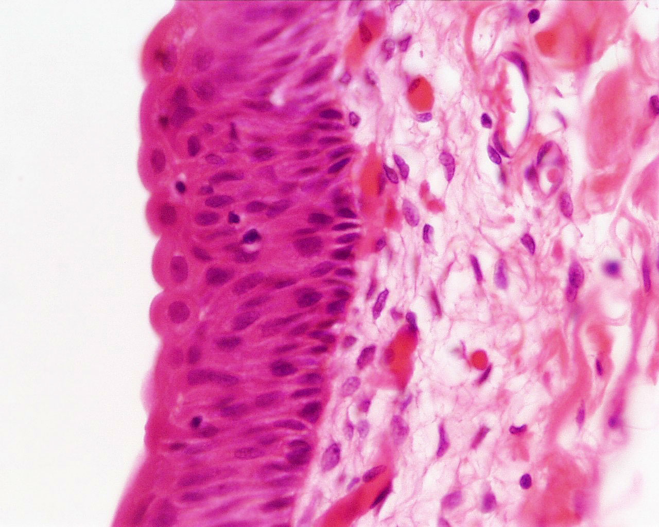
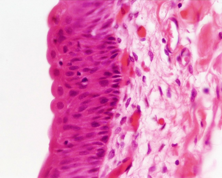
Urinary Bladder Histology
- primate bladder
- transitional epithelium (urothelium)
- Stain: H&E (x40)
- Renal Histology: Histology | Histology Stains | Renal Development
- Kidney - Nephron overview | Glomerulus | Vascular and renal poles | Medullary ray | tubules
- Ureter - Ureter labeled | Ureter epithelium
- Bladder - overview | wall 1 | wall 2 | transitional epithelium | Urinary Bladder Development
Links: Histology | Histology Stains | Blue Histology images copyright Lutz Slomianka 1998-2009. The literary and artistic works on the original Blue Histology website may be reproduced, adapted, published and distributed for non-commercial purposes. See also the page Histology Stains.
Cite this page: Hill, M.A. (2024, April 20) Embryology Bladder histology 003.jpg. Retrieved from https://embryology.med.unsw.edu.au/embryology/index.php/File:Bladder_histology_003.jpg
- © Dr Mark Hill 2024, UNSW Embryology ISBN: 978 0 7334 2609 4 - UNSW CRICOS Provider Code No. 00098G
Original file name: Bla42he.jpg
File history
Click on a date/time to view the file as it appeared at that time.
| Date/Time | Thumbnail | Dimensions | User | Comment | |
|---|---|---|---|---|---|
| current | 12:16, 30 August 2011 | | 1,280 × 1,024 (229 KB) | S8600021 (talk | contribs) | ==Urinary Bladder Histology== * primate bladder * transitional epithelium (urothelium) * Stain: H&E (x40) {{Renal Histology Links}} {{Blue Histology}} Original file name: Bla42he.jpg Category:Histology Category:Renal |
You cannot overwrite this file.
File usage
The following 4 pages use this file: