File:Bailey307.jpg

Original file (832 × 833 pixels, file size: 128 KB, MIME type: image/jpeg)
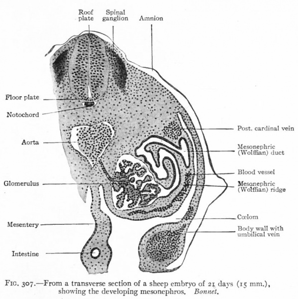
Fig. 307. From a transverse section of a sheep embryo of 21 days (15 mm)
Showing the developing mesonephros.
Bonnet.
After the tubules are formed, other condensations of the mesenchyme appear near their inner ends. A branch from the aorta enters each condensation and breaks up into a number of smaller vessels which ramify inside, the entire structure thus becoming a glomerulus. Each glomerulus pushes against the corresponding tubule, the latter becoming flattened and then growing around the glomerulus. In this way the glomerulus becomes surrounded by two layers of epithelium, except at the point where the vessels enter, and the whole structure the Malpighian corpuscle resembles very closely a renal corpuscle of the adult kidney. Waste products are removed from the blood through the agency of the glomeruli and are carried to the ducts by the mesonephric tubules (Fig. 307). The tubules themselves increase in length and become much coiled. Secondary and tertiary tubules also develop and become branches of the primary. Whether these develop from condensations of the mesenchyme or as buds from the primary tubules has not been determined. Each tubule consists of two parts (1) a dilated part around the glomerulus, composed of large flat cells and forming Bowman's capsule, and (2) a narrower coiled part leading from the glomerulus to the duct and composed of smaller cuboidal cells (Fig. 307).
- Text-Book of Embryology: Germ cells | Maturation | Fertilization | Amphioxus | Frog | Chick | Mammalian | External body form | Connective tissues and skeletal | Vascular | Muscular | Alimentary tube and organs | Respiratory | Coelom, Diaphragm and Mesenteries | Urogenital | Integumentary | Nervous System | Special Sense | Foetal Membranes | Teratogenesis | Gallery of All Figures
| Historic Disclaimer - information about historic embryology pages |
|---|
 |
Reference
Bailey FR. and Miller AM. Text-Book of Embryology (1921) New York: William Wood and Co.
Cite this page: Hill, M.A. (2024, April 18) Embryology Bailey307.jpg. Retrieved from https://embryology.med.unsw.edu.au/embryology/index.php/File:Bailey307.jpg
- © Dr Mark Hill 2024, UNSW Embryology ISBN: 978 0 7334 2609 4 - UNSW CRICOS Provider Code No. 00098G
File history
Click on a date/time to view the file as it appeared at that time.
| Date/Time | Thumbnail | Dimensions | User | Comment | |
|---|---|---|---|---|---|
| current | 11:11, 25 January 2011 | | 832 × 833 (128 KB) | S8600021 (talk | contribs) |
You cannot overwrite this file.
File usage
The following 3 pages use this file: