File:Bailey252.jpg
Original file (904 × 690 pixels, file size: 166 KB, MIME type: image/jpeg)
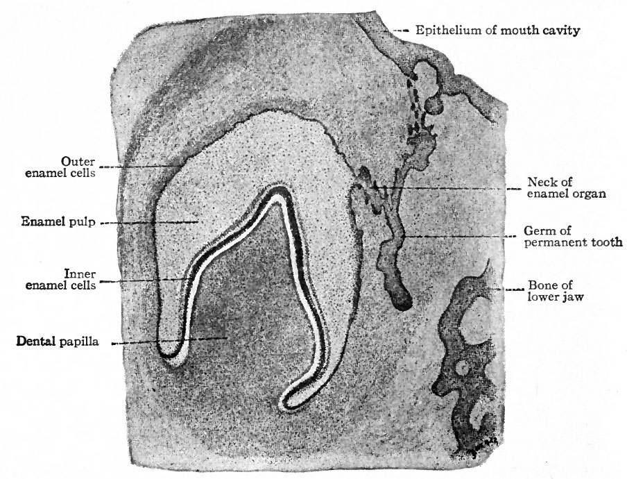
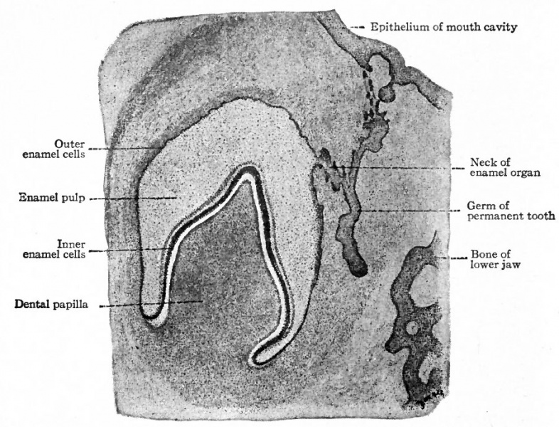
Fig. 252. Section of developing tooth from a 3 months human fetus
Szymonowicz.
The dental shelf is at first of uniform thickness, but in a short time five enlargements appear in it in each upper and lower jaw, indicating the beginnings of the milk teeth. When the embryo reaches a length of 40 mm. (an age of eleven to twelve weeks) the mesenchymal tissue on one side of these enlargements (above and to the inner side in the upper jaw, below and to the inner side in the lower jaw) becomes condensed and pushes its way into the epithelium. Each of these mesenchymal ingrowths is a dental papilla. Thus at this stage the anlage of each tooth is a mass of epithelium fitting cap-like over a mesenchymal papilla. The epithelium is the forerunner of the enamel organ; the papilla is destined to give rise to the dentine and pulp. The anlagen are connected with one another by intermediate portions of the dental shelf, and with the surface by the original ingrowth of epithelium.
- Text-Book of Embryology: Germ cells | Maturation | Fertilization | Amphioxus | Frog | Chick | Mammalian | External body form | Connective tissues and skeletal | Vascular | Muscular | Alimentary tube and organs | Respiratory | Coelom, Diaphragm and Mesenteries | Urogenital | Integumentary | Nervous System | Special Sense | Foetal Membranes | Teratogenesis | Gallery of All Figures
| Historic Disclaimer - information about historic embryology pages |
|---|
 |
Reference
Bailey FR. and Miller AM. Text-Book of Embryology (1921) New York: William Wood and Co.
Cite this page: Hill, M.A. (2024, April 16) Embryology Bailey252.jpg. Retrieved from https://embryology.med.unsw.edu.au/embryology/index.php/File:Bailey252.jpg
- © Dr Mark Hill 2024, UNSW Embryology ISBN: 978 0 7334 2609 4 - UNSW CRICOS Provider Code No. 00098G
File history
Click on a date/time to view the file as it appeared at that time.
| Date/Time | Thumbnail | Dimensions | User | Comment | |
|---|---|---|---|---|---|
| current | 10:35, 21 January 2011 | | 904 × 690 (166 KB) | S8600021 (talk | contribs) | {{Template:Bailey 1921 Figures}} Category:Human Category:Gastrointestinal Tract |
You cannot overwrite this file.
File usage
The following 2 pages use this file: