File:Bailey139.jpg
From Embryology

Size of this preview: 800 × 559 pixels. Other resolution: 961 × 671 pixels.
Original file (961 × 671 pixels, file size: 96 KB, MIME type: image/jpeg)
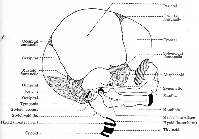
Fig. 139. Diagram of skull of new-born child
White areas represent bones of intramembranous origin; dotted areas represent bones (not derived from branchial arches) of intracartilaginous origin; black areas represent derivatives of branchial arches.
Combined from McMurrich and Kollmann.
- Text-Book of Embryology: Germ cells | Maturation | Fertilization | Amphioxus | Frog | Chick | Mammalian | External body form | Connective tissues and skeletal | Vascular | Muscular | Alimentary tube and organs | Respiratory | Coelom, Diaphragm and Mesenteries | Urogenital | Integumentary | Nervous System | Special Sense | Foetal Membranes | Teratogenesis | Gallery of All Figures
| Historic Disclaimer - information about historic embryology pages |
|---|
 |
Reference
Bailey FR. and Miller AM. Text-Book of Embryology (1921) New York: William Wood and Co.
Cite this page: Hill, M.A. (2024, April 25) Embryology Bailey139.jpg. Retrieved from https://embryology.med.unsw.edu.au/embryology/index.php/File:Bailey139.jpg
- © Dr Mark Hill 2024, UNSW Embryology ISBN: 978 0 7334 2609 4 - UNSW CRICOS Provider Code No. 00098G
File history
Click on a date/time to view the file as it appeared at that time.
| Date/Time | Thumbnail | Dimensions | User | Comment | |
|---|---|---|---|---|---|
| current | 12:34, 18 January 2011 | | 961 × 671 (96 KB) | S8600021 (talk | contribs) | ==Fig. 139. Diagram of skull of new-born child== White areas represent bones of intramembranous origin; dotted areas represent bones (not derived from branchial arches) of intracartilaginous origin; black areas represent derivatives of branchial arches. |
You cannot overwrite this file.
File usage
The following 3 pages use this file: