File:Bailey135.jpg
From Embryology
Size of this preview: 584 × 600 pixels. Other resolution: 940 × 965 pixels.
Original file (940 × 965 pixels, file size: 216 KB, MIME type: image/jpeg)
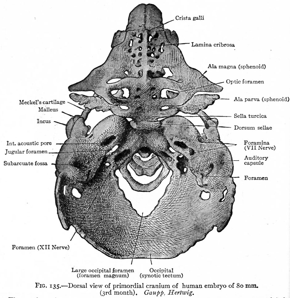
Fig. 135. Dorsal view of primordial cranium of human embryo of 80 mm
(3rd month).
The membrane bones of the roof of the skull have been removed. Through the large occipital foramen can be seen the first three cervical vertebrae.
Gaupp. Hertwig.
- Text-Book of Embryology: Germ cells | Maturation | Fertilization | Amphioxus | Frog | Chick | Mammalian | External body form | Connective tissues and skeletal | Vascular | Muscular | Alimentary tube and organs | Respiratory | Coelom, Diaphragm and Mesenteries | Urogenital | Integumentary | Nervous System | Special Sense | Foetal Membranes | Teratogenesis | Gallery of All Figures
| Historic Disclaimer - information about historic embryology pages |
|---|
 |
Reference
Bailey FR. and Miller AM. Text-Book of Embryology (1921) New York: William Wood and Co.
Cite this page: Hill, M.A. (2024, April 20) Embryology Bailey135.jpg. Retrieved from https://embryology.med.unsw.edu.au/embryology/index.php/File:Bailey135.jpg
- © Dr Mark Hill 2024, UNSW Embryology ISBN: 978 0 7334 2609 4 - UNSW CRICOS Provider Code No. 00098G
File history
Click on a date/time to view the file as it appeared at that time.
| Date/Time | Thumbnail | Dimensions | User | Comment | |
|---|---|---|---|---|---|
| current | 13:49, 18 January 2011 | | 940 × 965 (216 KB) | S8600021 (talk | contribs) | ==Fig. 135. Dorsal view of primordial cranium of human embryo of 80 mm== (3rd month). The membrane bones of the roof of the skull have been removed. Through the large occipital foramen can be seen the first three cervical vertebrae. Gaupp. Hertwig. |
You cannot overwrite this file.
File usage
The following 2 pages use this file: