File:Bailey031.jpg
Original file (768 × 1,101 pixels, file size: 219 KB, MIME type: image/jpeg)
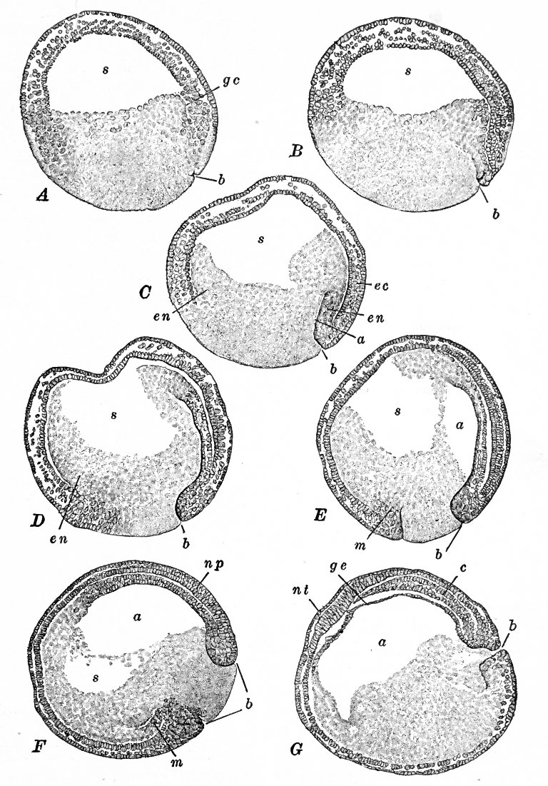
Fig. 31. Median sagittal sections showing successive stages of gastrulation in the frog's egg
Bracket, from Kellicott.
A, beginning of gastrulation
B, slight advance in invagination and beginning of epiboly
C, invagination and epiboly progressing, inflection of cells (involution) occurring around dorsal lip of blastopore which is now an obvious structure
D, epiboly has resulted in covering of a large part of yolk by lip of blastopore
E, blastopore is now circular and filled with the yolk plug (cf. Fig. 30, A, 4) and the archenteron appears as a small space
F, the blastoccel is nearly obliterated
G, gastrulation completed.
a, Archenteron; b, blastopore; c, rudiment of notocord; ec, ectoderm; en, entoderm; gc, gastrular cleavage, ge, entoderm (protentoderm) ; m, peristomal mesoderm; np, neural plate; w/, transverse neural ridge; s, blastocoel.
- Text-Book of Embryology: Germ cells | Maturation | Fertilization | Amphioxus | Frog | Chick | Mammalian | External body form | Connective tissues and skeletal | Vascular | Muscular | Alimentary tube and organs | Respiratory | Coelom, Diaphragm and Mesenteries | Urogenital | Integumentary | Nervous System | Special Sense | Foetal Membranes | Teratogenesis | Gallery of All Figures
| Historic Disclaimer - information about historic embryology pages |
|---|
 |
Reference
Bailey FR. and Miller AM. Text-Book of Embryology (1921) New York: William Wood and Co.
Cite this page: Hill, M.A. (2024, April 23) Embryology Bailey031.jpg. Retrieved from https://embryology.med.unsw.edu.au/embryology/index.php/File:Bailey031.jpg
- © Dr Mark Hill 2024, UNSW Embryology ISBN: 978 0 7334 2609 4 - UNSW CRICOS Provider Code No. 00098G
File history
Click on a date/time to view the file as it appeared at that time.
| Date/Time | Thumbnail | Dimensions | User | Comment | |
|---|---|---|---|---|---|
| current | 14:42, 17 January 2011 | | 768 × 1,101 (219 KB) | S8600021 (talk | contribs) | {{Template:Bailey 1921 Figures}} |
You cannot overwrite this file.
File usage
The following 3 pages use this file: