File:Adult skin histology 02.jpg

Original file (600 × 750 pixels, file size: 88 KB, MIME type: image/jpeg)
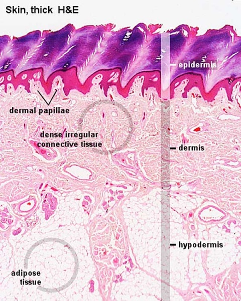
Adult Skin Histology
Histological section of skin showing the 3 main layers: epidermis, dermis and hypodermis layers. (Stain - Haematoxylin Eosin)
- epidermis - outer layer, stratified squamous epithelium.
Highlighted ringed regions show:
- dermis - middle layer, dense irregular connective tissue.
- hypodermis - inner layer, adipose tissue.
Note the junctional region between dermis and hypodermis contains macroscopically visible glands and blood vessels.
- Integument Histology Links: Adult Skin | Epidermis and Dermis | Thin Skin Epidermis | Thick Skin Epidermis | Elastic Fibres | Basal Cell Melanin | Foundations Practical Support | Integumentary System Development | Histology Stains
Links: Histology | Histology Stains | Blue Histology images copyright Lutz Slomianka 1998-2009. The literary and artistic works on the original Blue Histology website may be reproduced, adapted, published and distributed for non-commercial purposes. See also the page Histology Stains.
Cite this page: Hill, M.A. (2024, April 25) Embryology Adult skin histology 02.jpg. Retrieved from https://embryology.med.unsw.edu.au/embryology/index.php/File:Adult_skin_histology_02.jpg
- © Dr Mark Hill 2024, UNSW Embryology ISBN: 978 0 7334 2609 4 - UNSW CRICOS Provider Code No. 00098G
File history
Click on a date/time to view the file as it appeared at that time.
| Date/Time | Thumbnail | Dimensions | User | Comment | |
|---|---|---|---|---|---|
| current | 17:24, 14 February 2012 | | 600 × 750 (88 KB) | S8600021 (talk | contribs) | |
| 17:24, 14 February 2012 |  | 600 × 750 (108 KB) | S8600021 (talk | contribs) | {{Template:Blue Histology}} Category:Integumentary Category:Histology |
You cannot overwrite this file.
File usage
The following 9 pages use this file: