File:Absent cervical spine pedicle.jpg
Absent_cervical_spine_pedicle.jpg (600 × 338 pixels, file size: 30 KB, MIME type: image/jpeg)
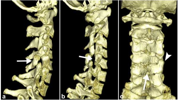
Absent Cervical Spine Pedicle
Volume Rendering Technique - Images of cervical spine. Lateral view VRT images illustrate the dorsally displaced right articular pillar of C5 (arrow in a).
The abnormal enlarged right intervertebral foramen of C4-5 (arrow in b) which is a consequence of the absent cervical pedicle is displayed on an oblique view VRT image.
Spina bifida occulta at the same level (arrow in c) as well as a reversed facet-joint on the right (arrowhead in c) are well depicted on a dorsal view VRT images (c).
Reference
<pubmed>21062465</pubmed>| BMC Med Imaging.
Copyright
© 2010 Guggenberger et al; licensee BioMed Central Ltd. This is an Open Access article distributed under the terms of the Creative Commons Attribution License (http://creativecommons.org/licenses/by/2.0), which permits unrestricted use, distribution, and reproduction in any medium, provided the original work is properly cited.
Original file name: Figure 3. 1471-2342-10-25-3.jpg
File history
Click on a date/time to view the file as it appeared at that time.
| Date/Time | Thumbnail | Dimensions | User | Comment | |
|---|---|---|---|---|---|
| current | 11:20, 21 December 2010 | | 600 × 338 (30 KB) | S8600021 (talk | contribs) | ==Absent cervical spine pedicle== Volume Rendering Technique - Images of cervical spine. Lateral view VRT images illustrate the dorsally displaced right articular pillar of C5 (arrow in a). The abnormal enlarged right intervertebral foramen of C4-5 (arrow |
You cannot overwrite this file.
File usage
The following 2 pages use this file: